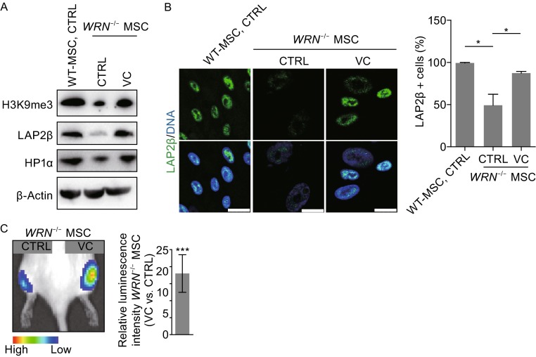
Figure 3.
VC restores epigenetic parameters and in vivo viability of WS MSCs. (A) Western blot analysis of the indicated proteins in MSCs. (B) VC increased heterochromatin markers by immunofluorescence staining. Representative immunofluorescence staining (left) and quantitative analysis (right) of LAP2β in vehicle or VC treated (7 days) WT MSCs and WRN -/- MSCs. Scale bar, 25 µm. (C) Luciferase activity of WS MSCs was detected by in vivo imaging system (IVIS) one week after implantation, and quantitative results were shown on the right. All data are represented as mean ± SEM. *P < 0.05, ***P < 0.001 by t test; n ≥ 3

