
Figure 3.
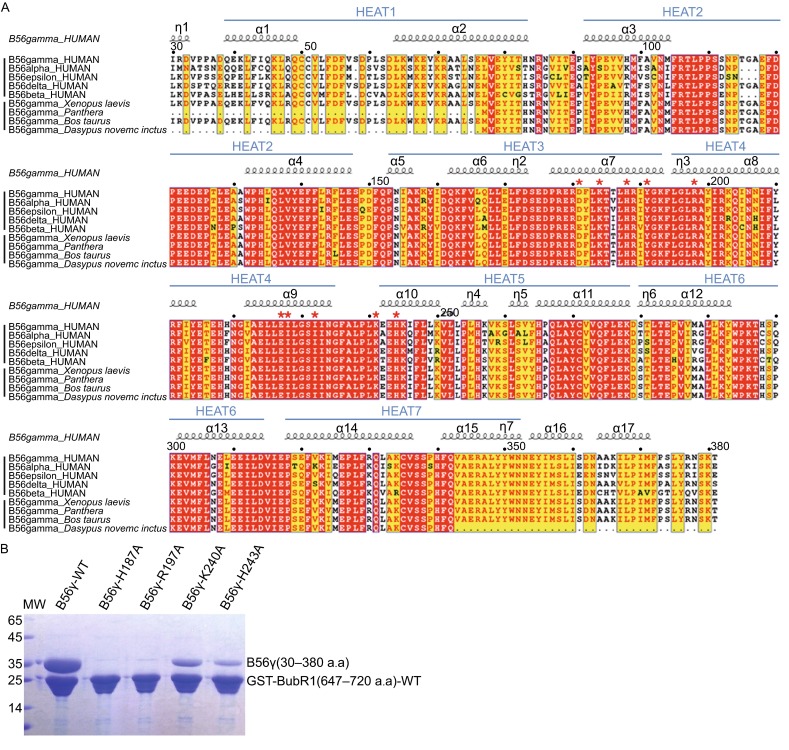
Sequence alignment of B56γ1 and mutagenesis of B56γ1. (A) B56 sequence alignment. The upper half shows the alignment of the five different human B56 isoforms, whereas the lower half demonstrates the sequence conservation of B56γ from different organisms. Numbers above indicate amino acid position based on the sequence of human B56γ1. B56γ has three isoforms with different C-terminal domains. Only B56γ1 is shown here. The strictly conserved residues are in white letters with red background and the conserved residues are in red letters with yellow background. Residues involved in BubR1 interaction are indicated with red asterisks. (B) Pulldown of mutant B56γ1 proteins by WT GST-BubR1(647–720).