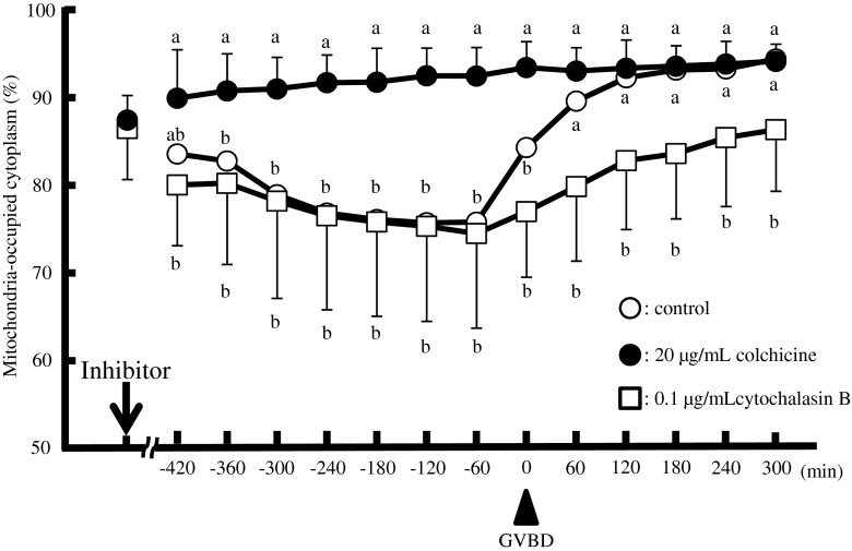
Fig. 5.
Time-dependent changes of the mitochondria-occupied area (percentage of total) in oocytes in the absence (open circles) or presence of colchicine (20 μg/mL, closed circles) or cytochalasin B (0.1 μg/mL, open squares). Data were compared among experiments with and without inhibitors and analysed by Fisher’s PLSD test following ANOVA (a–b, p < 0.01). The arrow and arrowhead indicate the time point when the inhibitors were added and the time of GVBD, respectively. Characteristics of oocytes used in this study were shown in Table 1

