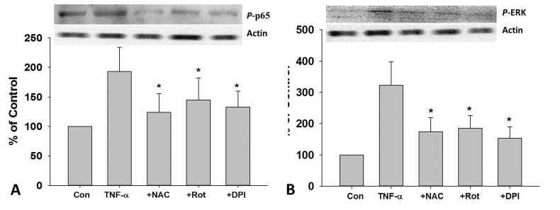
Fig. 5. Role of ROS in TNF-α-induced phosphorylation of NF-κB p65 and ERK in HUVECs.
After 2 h pretreated with DPI (10 MM), Rot (10 µM), NAC (1 mM), cells were treat ed with TNF-α (10 ng/mL) for 24 h, the protein expression of p-p65 (A) and p-ERK (B) was measured by Western blotting. Rot, rotenone; *p<0.05 vs. TNF-α treated group.

