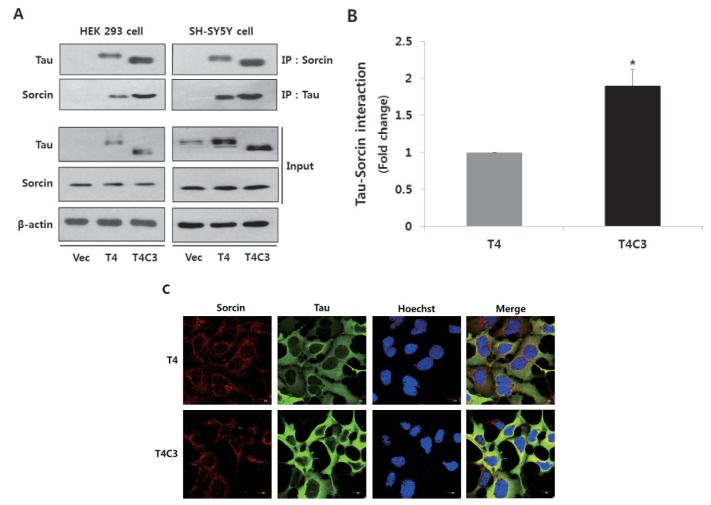
Fig. 1. Tau interacts with sorcin.
Representative images (A) and quantitative analysis (B) of immunoprecipitation of tau and sorcin. Intracellular localization of tau and sorcin (C). HEK 293 and SH-SY5Y cells were transfected with T4, T4C3, or vector. After 48 hr of transfection, cells were harvested. Cell lysates were immunoprecipitated with tau or sorcin antibody, and detected with Western blotting with respective antibodies. Vector was used for transfection control and agarose beads control in immunoprecipitation data to conform binding of agarose beads to tau or sorcin. Data were obtained from independent experiments (n=3) and expressed as mean±SD. *p<0.05 indicates significant differences between the indicated groups. To examine the intracellular localization of tau and sorcin, T4 or T4C3 transfected HEK 293 cells were stained with sorcin (red) or tau (green) antibody. Nuclei were visualized by Hoechst staining (blue). Scale bar 10 µm. Vec stands for vector (pcDNA3.1-) and IP for immunoprecipitation.

