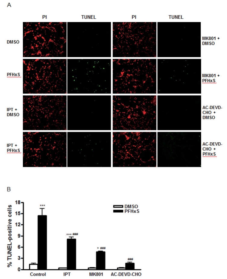
Fig. 2. Effects of imperatorin on CGC apoptosis induced by PFHxS.
Cells were pretreated with imperatorin (500 nM), MK801 (1 µM) or AC-DEVE-CHO (10 µM) for 1 h and treated with either PFHxS (300 µM) or DMSO as a vehicle control for 3 h. Then, cells were incubated in fresh media for 21 h. The apoptotic cells were stained with TUNEL (green) and all cells were counterstained with PI (red). (A) TUNEL- and PI-positive cells were monitored by fluorescence microscopy. Representative microscopic images from three independent experiments are presented (magnification, x200). (B). TUNEL– and PI-positive cells were counted. The number of TUNEL-positive cells was expressed as a percentage of the total number of cells. Data (% TUNEL-positive cells) are mean±SEM of three independent experiments. *p<0.05, ***p<0.001 vs. DMSO. ###p&0.001 vs. corresponding Control-treated cells (IPT, imperatorin).

