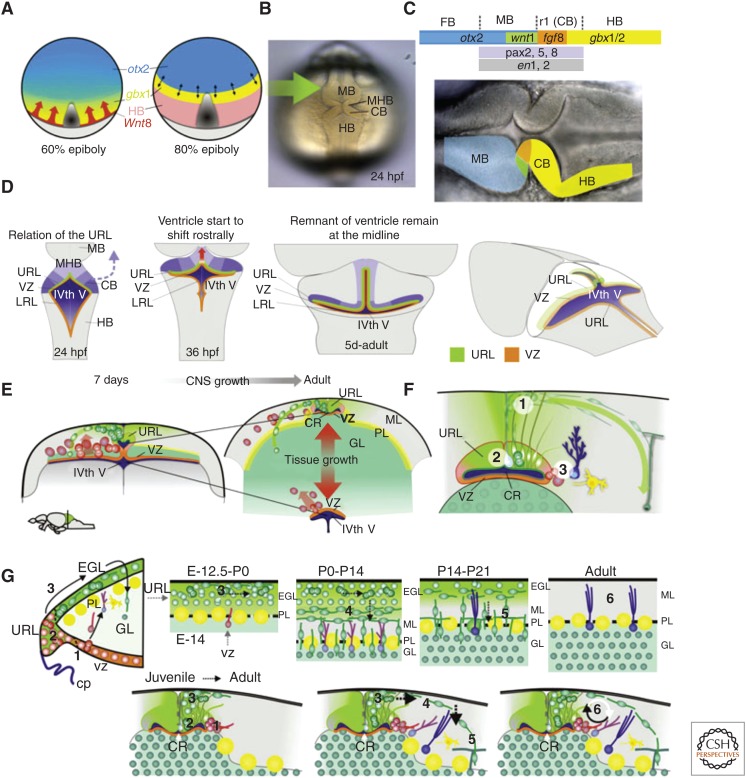
Figure 5.
Development and neurogenesis in the cerebellar niche. Cerebellar development in vertebrates depends on (A) the isthmic organizer positioned at the midbrain–hindbrain boundary (MHB) in the neural plate of zebrafish at the interface between otx (blue) and gbx (yellow) expression domains occuring during gastrulation, under control of Wnt8 secreted by the blastoderm margin (red arrows). Gray area 1/4 developing axial mesoderm. Light gray 1/4 yolk. (Panel A modified from data in Rhinn et al. 2005.) (B,C) Morphogenetic events of the cerebellar primordium. At 24 hpf, the MBH, midbrain structures, and the cerebellar primordium are clearly distinguishable in the zebrafish embryo. (C) At the end of gastrulation, a complex genetic network with several region-specific transcription factors and the secreted molecules Wnt1 and Fgf8 are expressed at the otx/gbx interface. The secreted signals from the isthmic organizer in turn determine the development of the surrounding mid- and hindbrain tissue. (D) Summary of the early morphogenetic events and the establishment of the progenitor domains. There is a morphogenetic rotation of the cerebellar primordium (blue arrow). From 36 hpf onward, the dorsomedial part of the fourth ventricle is shifted anteriorly creating a dorsomedial extension of the fourth ventricle (red arrow). Ventricularly located progenitors (orange line) are found in the lower rhombic lip (LRL) of the hindbrain and in the ventricular zone (VZ) of the cerebellum. Cerebellar progenitors adjacent to the roof plate are induced to turn into granule cell progenitors (upper rhombic lip [URL] green line). (E) Schematic summary of tissue growth and displacement of the progenitor niche. Displacement of the URL progenitor niche through tissue growth begins ∼7 dpf. During juvenile stages, there is vast generation of granule cells and massive expansion of the granule cell layer. The expanding granule cell layer separates the dorsomedial part of the IVth ventricle and the progenitors in the URL (green) from the rest of the IVth ventricle and the VZ. The addition of axonal fiber mass to the molecular layer further separates the URL progenitors dorsally from the roof plate/meninge. URL progenitors (green) are maintained dorsal to the cerebellar recessus in adult zebrafish, whereas ventricular-zone-derived progenitors and glia (orange) are found ventral to the recessus around the IVth ventricle. (F) Schematic summary of the zebrafish cerebellar progenitor niche. Neural progenitors derived from the URL are maintained in the dorsomedial part of the cerebellum around a remnant of the IVth ventricle (the cerebellar recessus). These progenitors give rise to granule neurons in a distinct outside-in fashion. Steps are numbered: (1) Polarized neuroepithelial-like progenitors (green) are restricted to the midline of the dorsal cerebellum. The progenitors give rise to rapidly migrating granule precursors (dark green) that initially migrate dorsolaterally. During this initial phase, the granule precursors still may proliferate. After reaching the meninge, the granule precursors change to a unipolar morphology and migrate in a ventrolateral direction toward the granule layer (GL). The granule precursors migrate into the GL and differentiate into granule neurons. (2) A few glia with a radial morphology (light blue), found close to the midline, are used as scaffolds during the initial dorsal migration of granule precursors. (3) Bergmann glia-like cells are interspersed in the Purkinje cells layer (PL) (dark blue). A low amount of Bergmann glia-like cells and inhibitory neurons (yellow) are generated from VZ progenitors that are found lateral and ventral to the progenitor niche. VZ progenitors are also found ventrally around the IVth ventricle (see E). (G) Comparison of the life-long maintained neurogenic program in the zebrafish cerebellum and the cerebellar developmental program in mammals. Numbered steps: (1) Ventricularly located VZ progenitors (orange) generate precursors (red) for inhibitory neurons and glia (yellow and blue). (2) In mammals, progenitors (green) in the URL feed the EGL with granule cell precursors. In zebrafish, no distinct EGL is discernable, although precursors do divide en route. (3) In mammals, greatly amplifying granule cell precursors are found in the EGL. The granule cell precursors are initially tangentially migrating. In contrast, a low level of amplification takes place in the zebrafish brain. (4,5) Granule cell precursors become postmitotic, differentiate, and migrate ventrally into the GL. (6) In the mammalian cerebellum, the URL and EGL are exhausted of their progenitors, whereas in zebrafish, primary URL progenitors and some VZ progenitors are maintained in a dividing state by a specialized niche. CB, cerebellar primordium; HB, hindbrain GL, granule cell layer; IVth V, fourth ventricle; LRL, lower rhombic lip; MHB, midbrain–hindbrain boundary; ML, molecular layer; PL, Purkinje cell layer; URL, upper rhombic lip; VZ, ventricular zone; CP, choroid plexus; CR, cerebellar recessus;, EGL, external granule layer. (From data in Kaslin and Brand 2012 and Kaslin et al. 2009, 2013; adapted, with permission, from the authors.)

