Figure 5.
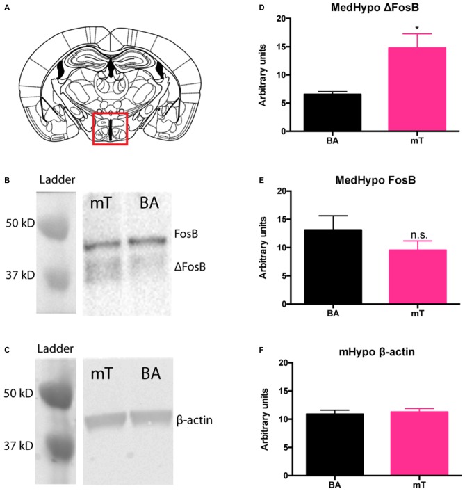
ΔFosB levels are increased in the medial hypothalamus (mHyp) following repeated mT exposure. Western blots for FosB and β-actin (loading control) were carried out on fresh-frozen brain tissue dissected from the mHyp (A). Protein bands were distinguished by size (B,C). Band intensities were calculated using ImageJ gel analysis tools. ΔFosB protein levels were significantly elevated (D) in the mHyp following 6 weeks of daily mT exposure compared to BA-exposed mice. Neither FosB levels (E), nor β-actin (F) differed between scent groups. All error bars shown represent SEM.

