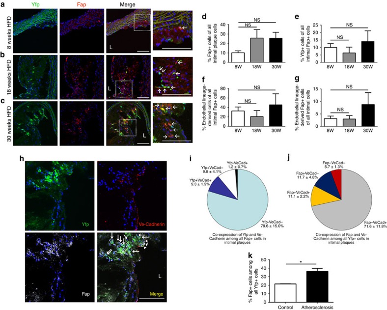
Figure 2. EndMT gives rise to fibroblast-like cells in intimal plaques.
(a–c) Immunofluorescence confocal microscopy of thoracic aortic sections from tamoxifen-induced end.SclCreERT;R26RstopYfp;ApoE−/− mice after (a) 8, (b) 18, (c) 30 weeks of HFD revealed Yfp+ cells co-expressing fibroblast-specific marker Fap within intimal plaques. L=lumen; scale bars, 100 μm. Insets at higher magnification as indicated. Arrows indicate co-positive cells. (d) Cell quantitation in intimal plaques revealed that after 8, 18 and 30 weeks of HFD, 10.2±2.1%, 25.7±9.0% and 25.6±6.3% (respectively) of cells expressed Fap, indicative of a fibroblast phenotype. (e) Quantitation (not accounting for the proportion of endothelial cells expressing Yfp) identified that at the same time-points, 10.0±2.6%, 6.4±3.9% and 14.0±7.2% (respectively) of intimal Fap+ cells co-expressed Yfp. (f) After accounting for the efficiency of our endothelial lineage tracking system (% of endothelial cells expressing Yfp), at the same time-points, we determined that 32.5±8.5%, 20.7±12.6% and 45.5±23.3% of intimal Fap+ cells were endothelial lineage-derived. (g) Again taking into account the efficiency of our endothelial lineage tracking system, at the same time-points, respectively, we determined that 3.1±1.0%, 3.0±1.4% and 8.8±4.7% of all intimal cells were endothelial lineage-derived Fap+ cells. For Fig. 1e–h staining was performed with at least four images evaluated from each of at least three spatially separated thoracic aortic sections per mouse (n=5 mice for 8 and 18 weeks; n=4 mice for 30 weeks). Data were averaged per animal then used for statistical analyses (one-way ANOVA). NS, not significant. (h) Four colour immunofluorescence confocal microscopy of thoracic aortic sections from tamoxifen-induced end.SclCreERT;R26RstopYfp;ApoE−/− mice after 18 weeks HFD demonstrating Yfp+Fap+Ve-Cadherin+ (arrow heads) and Yfp+Fap+Ve-Cadherin− cells (arrows). L=lumen; scale bars, 100 μm. Quantitation of Yfp+, Fap+ and Ve-Cadherin+ (VeCad) cells in intimal plaques as shown in h was performed (n=5 mice) to assess (i) the relative co-expression of Yfp and Ve-Cadherin among all Fap+ cells (j) the relative co-expression of Fap and Ve-Cadherin among all Yfp+ cells. (k) Fap expression was assessed among Yfp+ cells by FACS revealing that in control non-atherosclerotic end.SclCreERT;R26RstopYfp mice receiving chow diet, 21.5±0.2% of Yfp+ cells expressed Fap, while in tamoxifen-induced end.SclCreERT;R26RstopYfp;ApoE−/− mice after 18 weeks of HFD, 36.2±3.6% of Yfp+ cells expressed Fap. All mice studied in panel k were of the same overall age (24 weeks). *P<0.05, n=3 per group.

