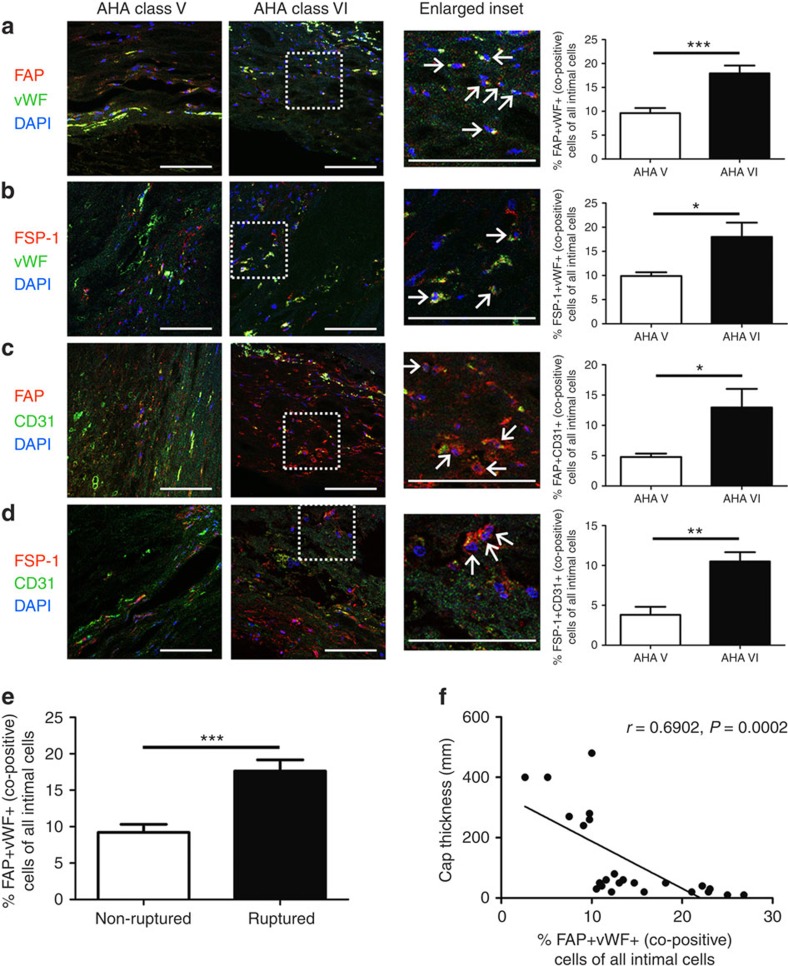
Figure 5. EndMT occurs in human atherosclerosis and is common in complex plaques.
Plaques identified in human samples obtained at autopsy from the abdominal aorta were classified as AHA type V or VI according to standard criteria40. Staining was performed using various endothelial–mesenchymal marker combinations as indicated. The % of co-positive cells per × 20 field was expressed as a function of the total number of DAPI+ cells (per field) as follows: (a) FAP/vWF, (b) FSP-1/vWF, (c) FAP/CD31, (d) FSP-1/CD31. Scale bars, 100 μm. Insets are shown at higher magnification as indicated and arrows indicate co-positive cells. (e) Percentage of FAP+vWF+ co-positive cells presented according to ruptured versus non-ruptured plaque morphology. *P<0.05, **P<0.01, ***P<0.001. (f) Scatter plot and regression line for aortic plaque cap thickness versus % FAP+vWF+ co-positive cells. r=0.6902, P=0.0002. For FAP/vWF, 24 randomly selected plaques from 16 patients were assessed (11 type V, 13 type VI), while for other combinations a minimum of 10 plaques from at least 6 different patients was randomly included per analysis.

