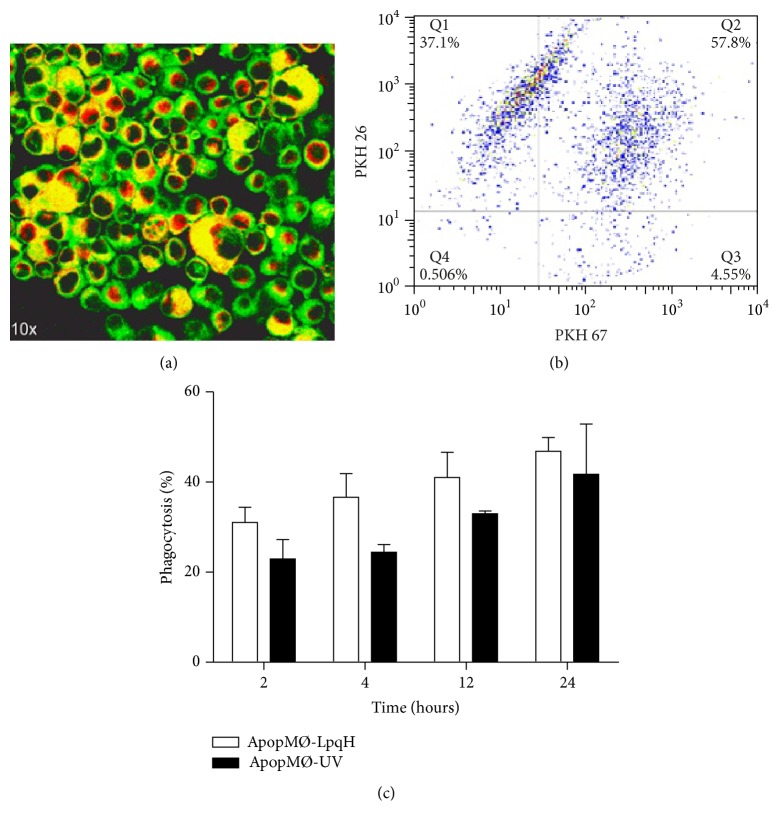
Figure 2.
Phagocytosis of apoptotic macrophages assessed by epifluorescence and flow cytometry. We conducted phagocytosis assays with J-774A.1 phagocytic cells labeled with PKH-67 (green fluorescence) and apoptotic MØs labeled with PKH-26 (red fluorescence). After two hours of incubation, in overlaid images, the confocal examination of mid-sectioned phagocytic cells showed enlarged MØs containing abundant yellow-fluorescent apoptotic bodies ((a), original 40x). Engulfed whole cells were not observed, perhaps due to their degradation within phagolysosomes. A representative dot blot shows double-labeled phagocytic cells in the upper right quadrant (b). Phagocytosis was quantitated by flow cytometry (c). Time-dependent phagocytosis was observed; that is, greater levels of apoptosis were observed at 24 h (47.7%). The results of three independent experiments are shown.

