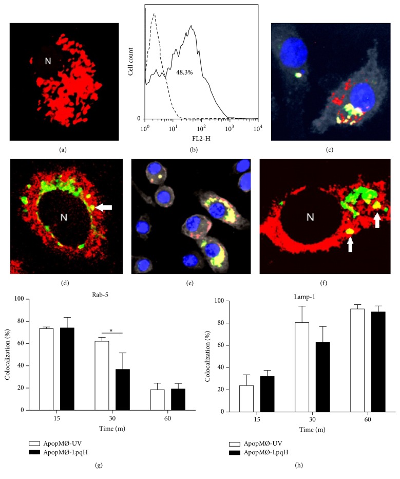
Figure 3.
Phagosomes with engulfed apoptotic macrophages mature into phagolysosomes. Phagocytosis assays were conducted with J-774A.1 cells and apoptotic MØs labeled with pHrodo, a marker that emits strong red fluorescence in an acidic environment. After 24 h of phagocytosis, confocal microscopy showed numerous pHrodo fluorescent apoptotic bodies in the majority of the cells ((a), original 100x). As determined by flow cytometry, 48.3% of the cells were found to be positive for pHrodo (b). To further determine phagosome maturation, phagocytic cells were cocultured for 15, 30, and 60 min with apoptotic MØs labeled green with PKH-67. The expression of the early endocytic marker Rab5 and LAMP1, a late endosome marker, was assessed by immunofluorescence. The cells were permeabilized and incubated with Cy5-labeled antibodies to Rab5 or LAMP1. In overlaid images, epifluorescence ((c), original 60x) and confocal microscopy ((d), original 100x) show cells containing yellow-fluorescent vesicles, indicating the colocalization of Rab5 with PKH-67-labeled apoptotic bodies. The colocalization of LAMP1 and engulfed apoptotic MØs was also demonstrated (e and f). As expected, the recruitment processes of Rab5 and LAMP1 followed inverse time courses (g and h). The results of three independent experiments are shown. Student's t-test was used to assess the statistical significance. ∗ p < 0.05. N, nucleus.

