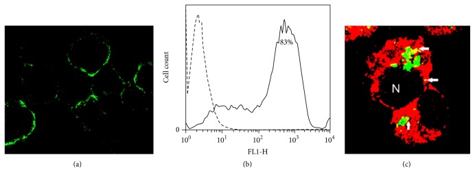
Figure 4.
Immunofluorescence demonstration of the role of the mannose receptor in the phagocytosis of apoptotic cells. To determine the expression of the MR on J-774A.1 macrophage-like cells, the cells were incubated with a Cy5-labeled anti-MR antibody. Confocal microscopy revealed patchy membrane fluorescence in many of the cells ((a), original 40x). A representative flow cytometry histogram demonstrated 83% MR expression (b); an isotype control is shown (dashed line). In overlaid confocal microscopy images, the role of the MR in phagocytosis was shown by the yellow fluorescent colocalization (arrows) of the MR (red fluorescence) with apoptotic bodies (green fluorescence) ((c), original 100x).

