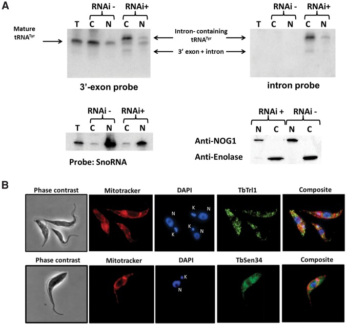
FIGURE 6.
Subcellular localization of tRNATyr, TbTrl1, and TbSen34. (A) RNA from total (T), nuclear (N), and cytoplasmic (C) fraction of 4 d induced (+) and uninduced (−) TbTrl1 RNAi cells was extracted and analyzed by Northern blot using a 3′ exon or intron probe. The intron-containing, mature tRNATyr and the 3′-exon–intron species are indicated by arrows. SnoRNA probe was used as control for nuclear fraction purity. The purity of the fractions was confirmed by Western blotting using anti-NOG1 (specific for nucleus) and anti-enolase antibodies (specific for cytoplasm). (B) Immunolocalization of His-tag TbTrl1 and Protein C-TbSen34. Anti-His-Tag and Anti-Protein C were used as primary antibodies for His-Tag TbTrl1 and detection Protein C-TbSen34, respectively. MitoTracker red was used as a mitochondrial marker, while DAPI was used to mark the position of nuclear (N) and mitochondrial DNA (kinetoplast) (K). TbTrl1 and TbSen34 are shown in green. A background was added to the TbSen34 images in order to get a more harmonious figure.

