Figure 1.
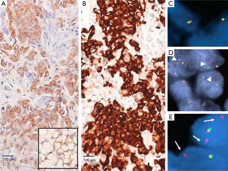
Different ALK staining expression at IHC with clone 5A4 (A) and Ventana D5F3 CD assay (B). Insert in image (A) shows the ALK positivity in signet ring tumor cells with a thin rim of brownish-stained cytoplasm dislocated under the nuclear membrane. FISH analysis showing tumor cells with normal ALK set-up with one normal fusion signal and one single red/orange signal (C); ALK positivity by gene deletion with single 3' orange rearranged signals (deleted green signal) (D, arrowheads) and ALK positivity by inversion with “broken apart” signals, 2 or more signal diameters apart (E, arrows). IHC, immunohistochemistry; ALK, anaplastic lymphoma kinase; FISH, fluorescence in situ hybridization.

