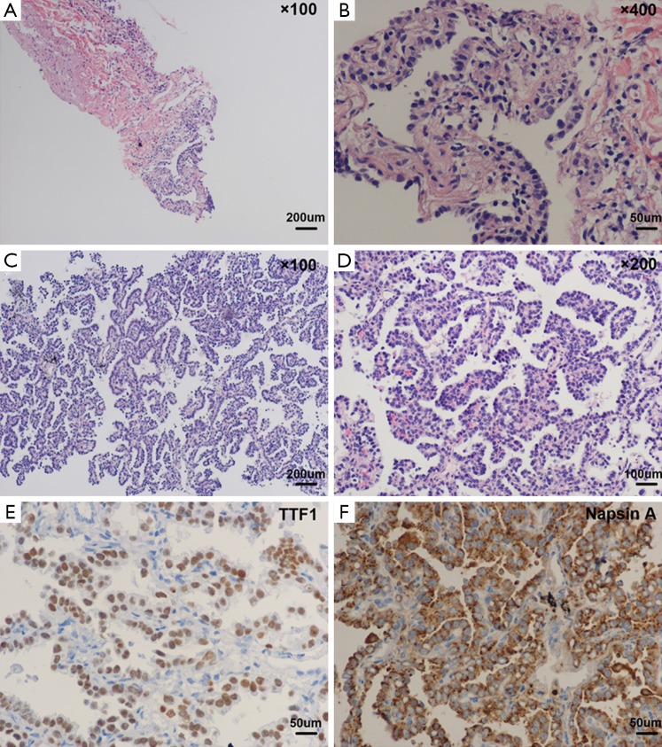
Figure 3.
Photomicrograph of the lung tissue from biopsies. (A,B) Biopsy performed on April 17, 2014 showed small pieces of lung tissue showed inflammatory fibroplasias, occasionally glandular structures could be found with mild to moderate atypia; (C,D) H & E staining of biopsy performed on August 14, 2014 reported high differentiation adenocarcinoma; (E,F) IHC staining of TTF1 and Napsin A. IHC, immunochemistry; TTF1, thyroid transcription factor-1.

