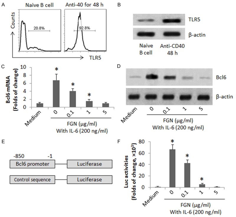
Figure 1.

FGN suppresses Bcl6 expression in B cells. CD19+ CD138- B cells were isolated from the spleen of naïve mice by MACS. (A, B) The naive B cells were cultured for 48 h in the presence of anti-CD40 Ab (20 ng/ml). The gated histograms indicate the frequency of TLR5+ B cells (A). The immune blots indicate the TLR5 protein in the B cell extracts (B). (C, D) The naive B cells were cultured in the presence of IL-6 and anti-CD40 with or without the presence of FGN (at graded concentrations) for 48 h. The cell extracts were analyzed by RT-qPCR and Western blotting. The bars indicate the mRNA levels of Bcl6 (C). The immune blots indicate the protein levels of Bcl6 (D). (E) A sketch of Bcl6 promoter reporter gene. (F) The bars indicate the Bcl6 promoter activities. The data of bars are presented as mean ± SD. *, p<0.01, compared with the medium group. The data are representatives of 3 independent experiments.
