Figure 1.

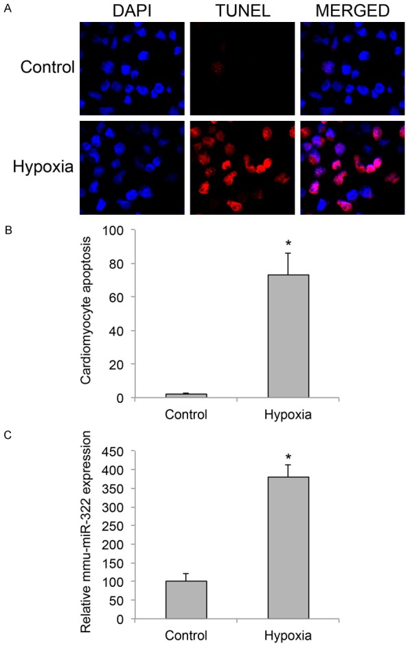
Cardiomyocyte apoptosis and miR-322 upregulation under hypoxia condition. Murine neonatal cardiomyocytes (postnatal 1~2 days old) were cultured in 48-well plates (3,000 cells/well) and treated with a gas mixture of 95% N2 and 5% CO2 for 2 h to induce hypoxia. A. 24 h after hypoxia treatment, a TUNEL assay (Red) was carried out to examine apoptosis in cardiomyocytes. Cardiomyocytes were identified by DAPI nuclear staining (Blue). B. The averaged percentages of apoptotic cardiomyocytes (DAPI/TUNEL (%)) were compared between control and hypoxia cultures (*P < 0.05). C. 24 h after hypoxia treatment, relative expression levels of mmu-miR-322 in cardiomyocytes were compared between control and hypoxia conditions (*P < 0.05).
