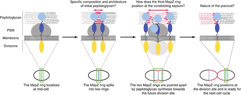
Figure 7. Model of MapZ positioning at the division site.
The upper part of the figure shows magnifications of the different cell division stages of the pneumococcus that are presented in the lower part. MapZ localizes at the interface between the two cell halves of newborn cells. Black and grey ovals distinguish the cell wall of each half-cell. The cytoplasmic domain of MapZ, MapZextra1 and MapZextra2 are represented by yellow, dark and light blue ovals, respectively. Initial peptidoglycan synthesis (red ovals) is recognized by MapZextra2 that is shifted towards the cell equator of the two daughter cells. Insertion of peptidoglycan allowing cell elongation and whose composition might be different from that of initial peptidoglycan is shown as open red ovals. The cytoplasmic part of the divisome and the peptidoglycan synthesis machinery (PSM) are shown in light and dark grey, respectively. For visual clarity, the divisome is not indicated in the first two stages of the cell division process. The question mark indicates the possible structural rearrangement of MapZextra when localizing as a third ring at the constricting septum.

