
Figure EV3. Myofibroblasts express UPR markers during skin fibrosis.
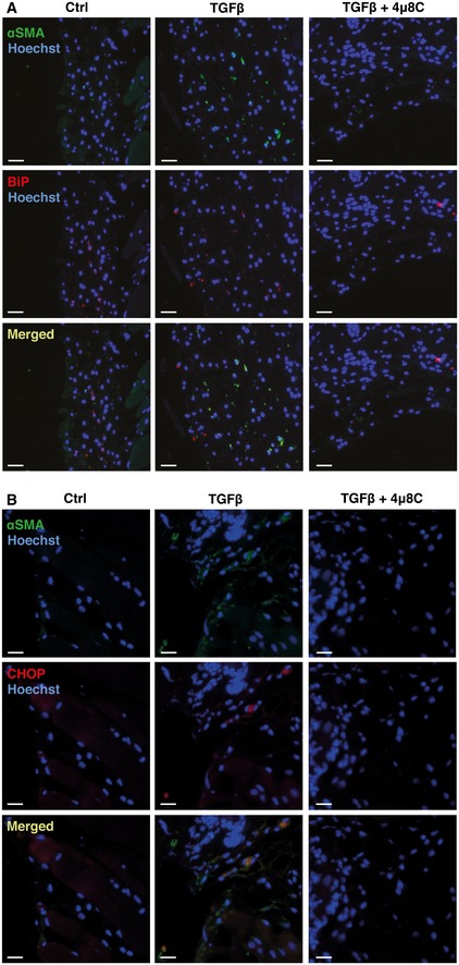
- Representative images of skin from control or 4μ8C‐treated C57BL/6 mice with TGFβ‐induced skin fibrosis. Sections of skin were co‐stained with anti‐αSMA and anti‐BiP antibodies. Scale bars = 50 μm.
- Representative images of mouse skin co‐stained with anti‐αSMA and anti‐CHOP antibodies. Scale bars = 50 μm.