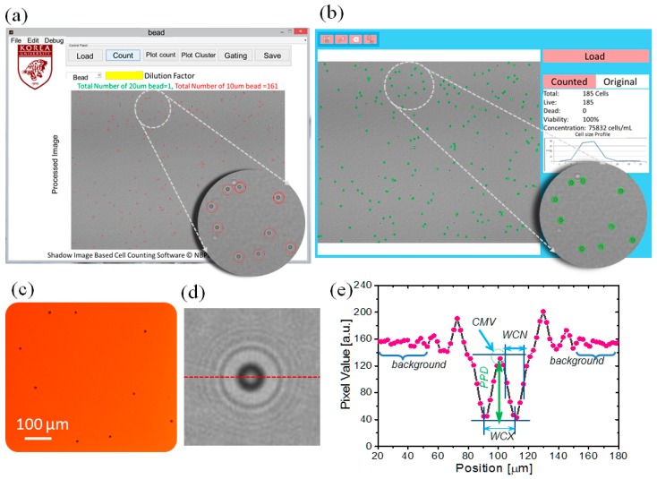
Figure 3.
Snapshot of the custom-developed application software. (a) Snapshot of custom-developed Windows application software; (b) snapshot of custom-developed Android application; (c) optical micrograph of region of interest at 100× magnification; (d) diffraction pattern of a single microparticle; (e) intensity profile of the diffraction pattern in (d) explaining the custom-developed diffraction parameters along the dotted red line in (d).

