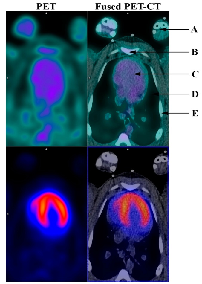
Figure 1.
Angiogenesis PET (RGD PET) (top row) and 82Rb dipyridamole stress PET (bottom row) before induced myocardial infarction. (A) Front limbs; (B) Sternum; (C) Heart; (D) Lungs; (E) Ribs. Angiogenesis is part of the healing process following an ischemic injury and is vital for the post-ischemic repair of the myocardium. It is associated with the remodeling of the left ventricle and thus prognosis following myocardial infarction [1]. Therefore, it is of particular interest to be able to noninvasively monitor angiogenesis. This might not only permit risk stratification of patients following myocardial infarction, but could also facilitate development and improvement of new therapies directed towards stimulation of the angiogenic response. During angiogenesis endothelial cells must adhere to one another to form new microvessels. This is a process modulated by the extracellular matrix including integrins. Specifically, αvβ3 integrin is highly expressed in activated endothelial cells and has been identified as a critical modulator of angiogenesis and is therefore a potential target for directly imaging angiogenesis [2,3]. Existing noninvasive imaging methods directed towards the evaluation of angiogenesis have however been somewhat limited, possibly due to the fact that myocardial angiogenesis following myocardial infarction might be focal and therefore difficult to detect. Furthermore, most of the previous studies in angiogenesis imaging have been performed in smaller animals, mostly rats [4,5,6,7,8,9,10,11,12,13]. 68Ga-NODAGA-E[c(RGDyK)]2 (RGD) has recently been developed by us as an angiogenesis positron-emission-tomography (PET) ligand targeted towards αvβ3 integrin [14]. In the present study, we induced myocardial infarction in Göttingen minipigs [15]. Successful infarction was documented by 82Rubidium (82Rb)-dipyridamole stress PET and computed tomography (CT) (Siemens mCT, Siemens, 128-slice CT, Knoxville, USA). RGD uptake was demonstrated in the infarcted myocardium one week and one month after induction of infarction by RGD-PET. The study was approved by the National Authority in Denmark (approval number: 2014-15-0201-00191). During the PET acquisition minipigs were anesthetized as described in detail previously [15]. Baseline 82Rb rest and stress myocardial perfusion were performed the week prior to induction of myocardial infarction as a 7 min dynamic PET myocardial perfusion rest scan under administration of 1000–1200 MBq 82Rb followed by a 7 min dynamic dipyridamole stress PET-CT. Dipyridamole (140 µg/kg/min) was given as a continuous intravenous infusion over 4 min prior to 82Rb-tracer injection 3–5 min after the completion of dipyridamole infusion. The RGD-PET was performed as a 10 min ECG-gated scan 45 min after administration of 100 MBq RGD. PET images were analyzed using Cedars-Sinai Cardiac Suite (Cedars-Sinai Medical Center, Los Angeles, CA, USA) for Syngo. Via (Siemens, Knoxville, TN, USA). The figure shows RGD and 82Rb stress PET images before induction of myocardial infarction. 82Rb stress PET showed even distribution of 82Rb in the left ventricle while the RGD PET showed no RGD uptake.

