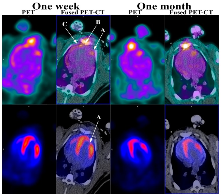
Figure 2.
RGD (top row) and 82Rb stress PET (bottom row) one week and one month after induced myocardial infarction. (A) Myocardial infarction; (B) Sternotomy; (C) Pericardium. As shown, the 82Rb stress PET (bottom row) showed a myocardial perfusion defect in the anterior wall of the left ventricle myocardium one week and one month after induced myocardial infarction confirming a myocardial infarction corresponding to an area supplied by the ligated branch from LAD. This myocardial perfusion defect was also present at rest (not shown). Furthermore, the RGD PET (top row) showed RGD uptake in the infarcted myocardium one week and one month following myocardial infarction. In addition, RGD PET showed RGD uptake in the sternum after sternotomy and pericardium, most likely due to the opening as part of the infarct induction procedure. As previously mentioned, most of the previous work in angiogenesis imaging have been done in smaller animals. The minipig heart and the human heart are very much alike, which makes the findings in this study even more encouraging and adds to the few, mostly very small, studies performed in human [16,17,18]. The perspectives are very intriguing and might permit the evaluation of new treatment strategies targeted towards increasing the angiogenetic response, e.g., stem-cell treatment.

