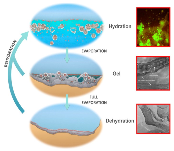
Figure 7.
Protocells cycling between three coupled phases. Hydrated phase (Top), inset: protocells containing DNA growing out of a dried mixture of DNA and phospholipid, hydrated by water, stained with acridine orange. Gel phase (Center), inset: hydrogel of lipid with approximately 50% water by weight showing lipid vesicles fusing into lamellae. Dehydrated phase (Bottom), inset: freeze fracture image of anhydrous lipid lamellae of phosphatidylcholine. Micrographs credit: David Deamer.

