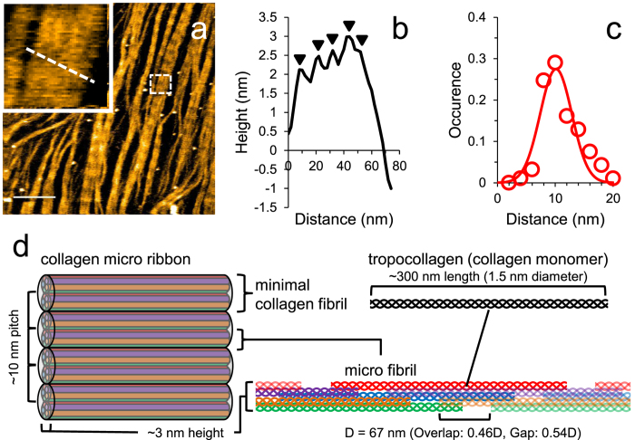
Figure 1. Collagen fibrils in a collagen microribbon on a HS-AFM stage.
(a) HS-AFM image of collagen microribbon assembled from type I collagen of rat tail. These images are taken from Supplementary Movie 1. The region highlighted by a dashed box is enlarged in the inset, where five minimum collagen fibrils lie vertically. Scanning rate, 10 s/frame; Scan area, 1000 × 1000 nm2 with 500 × 500 pixels; Bar, 200 nm; Z-scale, 5 nm. (b) Height profile along the dashed line in the inset of (a). The peaks are indicated by arrowheads. (c) Distribution of the distance between collagen fibrils in collagen microribbon. (d) Schematic view of hierarchical structure of collagen microribbon. Five triple helical collagen molecule units (red, blue, green, orange and purple) associate into a microfibril with gaps and overlaps. Two microfibrils form a minimal collagen fibril. From this scheme, the number of collagen monomers is estimated to be 2975 per square μm.

