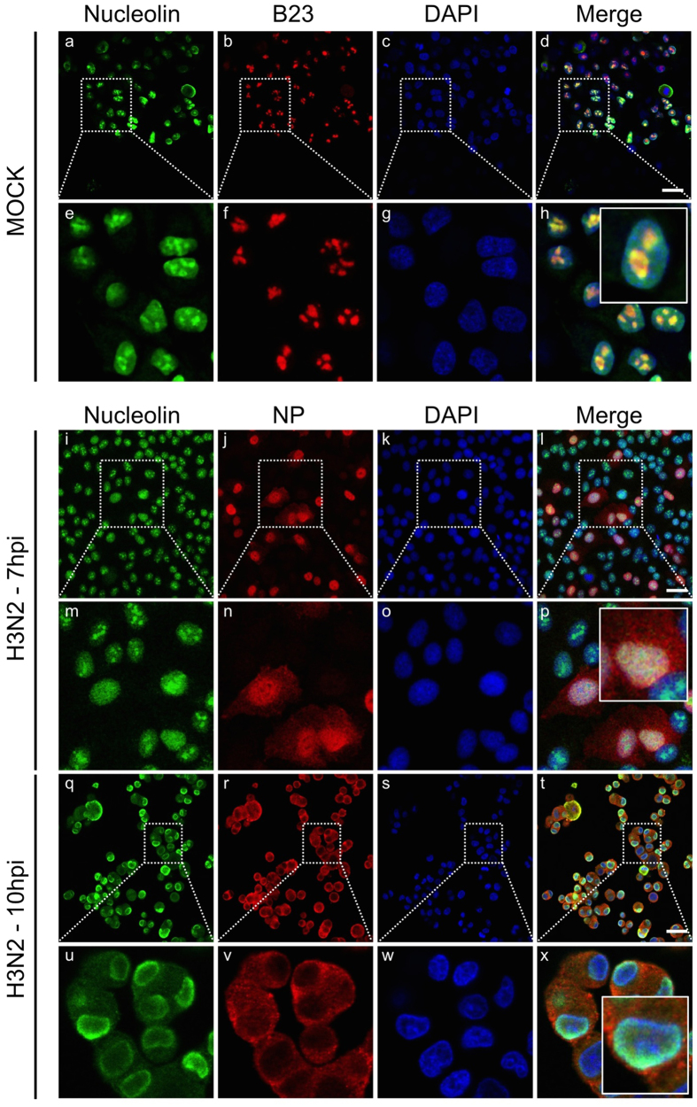
Figure 1. Host nucleolin is dynamically relocalized during the course of influenza infection.
Immunofluorescence staining of nucleolin (green) and B23 (red) in mock-infected cells (panels a to h) or NP (red) in A549 cells infected with H3N2 virus at a MOI of 1 (panels i to x) was performed at different times, as indicated. Nuclei were counterstained with DAPI (blue, panels c,g,k,o,s,w). Merged fluorescent signals are presented in panels d,h,i,p,t and x. Cell details are enlarged (inset). Scale bar = 10 μm.

