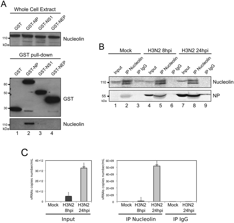
Figure 3.
NP interacts with endogenous nucleolin (A) A549 cells were transfected with plasmids coding GST or, alternatively, GST fused to NP, NS1 or NEP. Cell lysates were subject to a pull-down assay. Whole cell extracts and pull-down proteins were resolved on SDS-PAGE and immunoblotted with anti-GST and anti-nucleolin antibodies. Top-panel shows detection of the endogenous nucleolin within whole cell lysates. The bottom panel shows the level of expression of pulled-down GST fusion and endogenous nucleolin. (B) Cells were infected during 8 or 24 hours with H3N2 (MOI 2). Immunoprecipitations (IP) were performed on the whole cell extract with polyclonal anti-nucleolin or control IgG antibodies, as indicated. The presence of nucleolin and NP in immunopurified complexes was checked by western blot analysis. Whole cell extracts (Input) were used as control. (C) Cells were infected during 8 or 24 hours with H3N2, as before. Immunoprecipitations were performed with the corresponding whole cell extract proteins, using polyclonal anti-nucleolin or control IgG antibody. RNAs associated with purified complexes were extracted and M vRNAs were quantified by specific RT-qPCR (M vRNAs copies number/mL). Total RNAs were extracted from Mock- or H3N2-infected cells and used as controls (Input). ***P-value <0.001.

