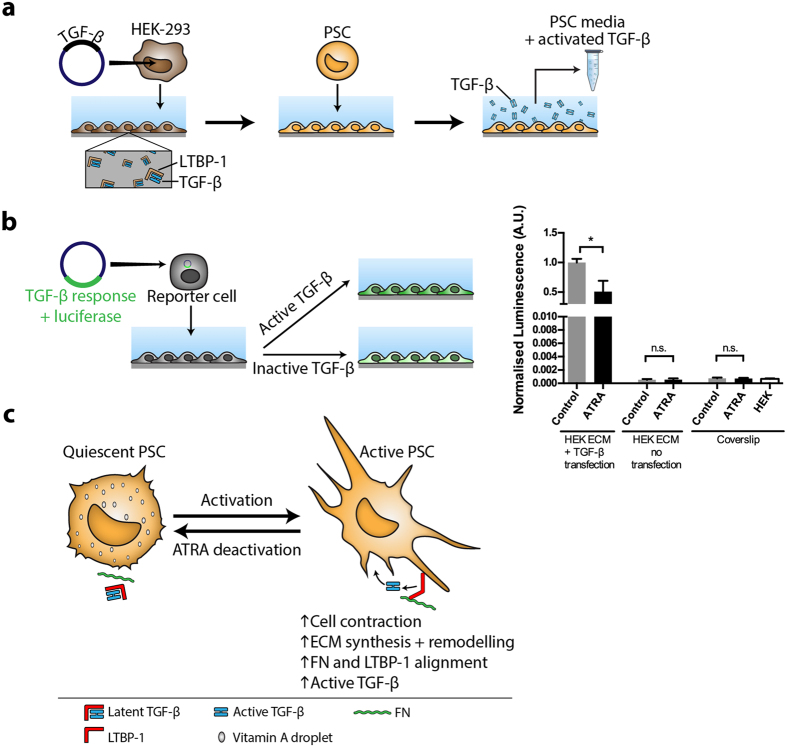
Figure 5.
The effect of ATRA treatment on liberation of TGF-β from its LTBP-1 latent form (a) Whole TGF-β transfected or non-transfected HEK-293 cells were cultured for 7 days and removed (first arrow). Treated or control PSCs were incubated on matrices for 48 hours during which they can release TGF-β from LTBP-1 into the culture media (second arrow). Active TGF-β containing conditioned culture media was collected. FN, fibronectin. (b) Left panel; schematic representation of TGF-β bioassay in which TGF-β reporter cells with a luminescence vector were incubated with active TGF-β containing control or ATRA treated PSC culture media. After ATRA treatment, TGF-β activation by PSCs was significantly reduced (right panel). There were no significant differences in luciferase signal between control and ATRA treated PSCs when cells were seeded on non-transfected HEK-293 ECM or on glass coverslips. The active TGF-β signals were negligible in the following cases: HEK-293 cells or PSCs seeded on glass and PSCs seeded on matrices previously deposited by non-transfected HEK-293. Representative of 3 independent experiments; *p < 0.05, two-tailed Student’s t-test, error bars show means ± SEM, A.U. Arbitrary units. (c) Summary schematic illustrating the effects PSC deactivation on ECM remodeling capacity.

