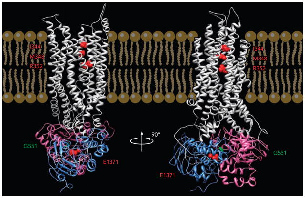
FIGURE 1. A modeled CFTR structure.
Side views of a homology model of CFTR based on the prokaryotic ABC exporter, Sav1866 (54). Gray, TMDs; purple, NBD1; blue, NBD2. Three residues in TM6 mentioned in this review (from top to bottom: I344, M348, R352) are labeled in red. G551 in NBD1 is labeled in green. E1371 in NBD2 is labeled in red (courtesy of Dr. David Dawson).

