Figure 1.
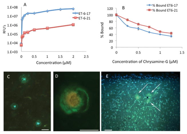
(A) Novel hydrophilic amyloid binding compounds ET6-17 and ET6-21 (LogP 1.80 and 1.06 respectively) bind to amyloid fibrils. N=3 at each point. (B) A competitive binding study holding the bulk phase concentration constant at 1.5 μM shows that binding is competitive with Chrysamine-G, consistent with specificity to the Thioflavin binding site. N=3 at each point. (C) ET6-21 specifically stains amyloid plaques (green/blue) in a section from the frontal cortex of an AD autopsy case (D) ET6-21 labels cerebral amyloid angiopathy (green) in the prefrontal aged dog brain (red/yellow labeling is due to blood cells inside the lumen of the blood vessel showing autofluorescence). Images were collected on an Olympus BX-51 epifluorescent microscope using the broadpass filter. (Scale bars represent 50 μm length). RFU: Relative Fluorescence Unit.

