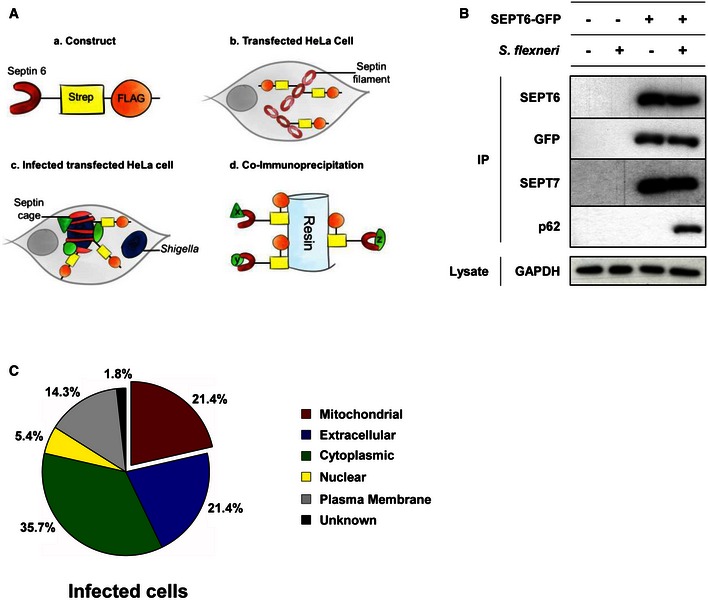
Figure 2. Proteins enriched at the Shigella–septin cage.

- Schematic representation of the proteomic experiments used to isolate proteins enriched at the Shigella–septin cage. (a) A SEPT6–STREP–FLAG construct was (b) transfected in HeLa cells for 24 h. (c) Cells were infected with Shigella flexneri AfaI for 4 h, harvested, and (d) SEPT6–STREP–FLAG cage‐associated proteins were isolated through co‐immunoprecipitation (Co‐IP).
- HeLa cells stably expressing SEPT6‐GFP were infected with S. flexneri AfaI for 4 h, harvested and then tested with Co‐IP. Empty vector and/or uninfected HeLa cells were used in parallel as control. GFP‐trap magnetic agarose beads were used to isolate SEPT6‐GFP from cells, and extracts were immunoblotted for SEPT6, GFP, SEPT7 or p62. The lysates were immunoblotted for GAPDH as control for cellular protein levels.
- The protein pulldown experiments identified 56 proteins putatively associated with the Shigella–septin cage and then categorised into six groups based on the analysis from the Gene Ontology database. See also Dataset EV1.
