
Figure EV3. Mitochondria support septin assembly into rings and cages.
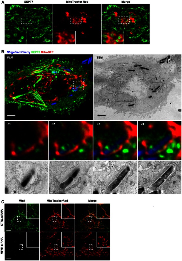
- HeLa cells were stained with MitoTracker Red CMXRos, treated with cytochalasin D for 30 min and then fixed and labelled for endogenous SEPT7 for confocal microscopy. Inset images highlight mitochondria closely associated with septin rings. The scale bar represents 5 μm.
- HeLa cells stably expressing SEPT6‐GFP were transfected with mito‐BFP for 24 h, infected with Shigella‐mCherry for 4 h 40 min and processed for CLEM. Z‐stack (Z1–Z4) series of septin cages were acquired by fluorescent light microscopy (FLM), and then samples were processed for TEM. SEPT6 is shown in green, mitochondria in red and Shigella in blue. The Z‐stack (Z1–Z4) series clearly shows the septin‐compartmentalised autophagosome and the mitochondrial membrane. The scale bar represents 5 μm.
- HeLa cells were treated with control (CTRL) or Mfn1 siRNA for 72 h, labelled with MitoTracker Red CMXRos, fixed for confocal microscopy and labelled with antibodies to Mfn1. The scale bar represents 5 μm. Inset images highlight Mfn1 associated with fused mitochondria fusion in CTRL cells, or absence of Mfn1 associated with fragmented mitochondria in Mfn1‐depleted cells.