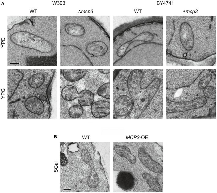
Figure EV1. Altered levels of Mcp3 have no effect on mitochondrial ultrastructure.

- WT and mcp3Δ cells in two different genetic backgrounds (W303 and BY4741) were grown in glucose (YPD) or glycerol (YPG) containing medium and analysed by thin‐section transmission electron microscopy. Representative images of mitochondria are shown. Scale bars, 200 nm.
- WT cells carrying an empty plasmid (WT) and cells overexpressing MCP3 from a plasmid under control of the TPI promoter (MCP3‐OE) were grown in galactose‐containing minimal medium (SGal) and analysed by thin‐section transmission electron microscopy. Representative images of mitochondria are shown. Scale bar, 200 nm.
