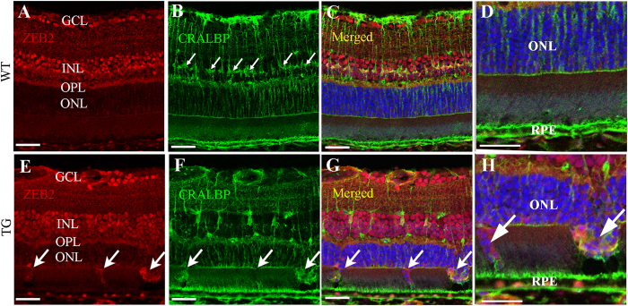
Figure 3. Increased immunoreactivity for ZEB2 in the outer retina after induced Müller cell disruption.
An antibody against CRALBP was used to stain Müller cells. (A–D) ZEB2 was expressed in the ganglion cell layer (GCL), the inner nuclear layer (INL) and the outer plexiform layer (OPL) but hardly detected in the outer nuclear layer (ONL) in the wild type (WT) retina. The small arrows in B point to Müller cell bodies in the INL. (E–H) Transgenic (TG) retina collected 3 months after induced Müller cell disruption. Increased immunoreactivity for ZEB2 was observed in the outer retina where Müller cell ablation resulted in defects is the outer limiting membrane (small arrows in E–G) through which degenerating photoreceptor cell bodies protruded into the subretinal space (large arrows in H). (D,H) are higher power images of the outer retina in (C,G) respectively. RPE = retinal pigment epithelium. Scale bars: 50 μm.

