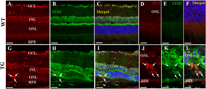
Figure 4. Double labelling for ZEB2 and isolectin B4 (IB-4) 3 months after Müller cell disruption.
(A–F) Wild type retinas. The normal retinal vasculature is distributed within the inner retina but absent in the outer retina. (G–L) Transgenic retinas collected 3 months after Müller cell disruption. Deep intraretinal neovascularisation that had developed 3 months after induced Müller cell disruption expressed ZEB2 (arrows in (G–L). (D–F,I–L) are higher power images of (A–C,G–I) respectively. GCL-ganglion cell layer, INL = inner nuclear layer, ONL = outer nuclear layer, RPE = retinal pigment epithelium. Scale bars: 50 μm in (A–C,G–I); 25 μm in (D–F,J–L).

