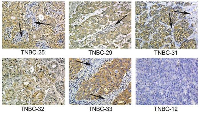
Figure 5.
Immunohistochemical Detection of α-Lactalbumin Protein in Human TNBC Tumors. Immunohistochemistry for α-lactalbumin performed on formalin-fixed paraffin embedded human TNBC whole tissue sections showing that 5/6 (83%) tumors showed cytoplasmic immunoreactivity that was diffuse throughout the carcinoma with varying levels of intensity ranging from weak (TNBC-32) to moderate (TNBC-33). Note that the background stromal cells (arrows) are negative. TNBC-12 is negative for protein expression of α-lactalbumin, correlating with the absent gene expression (see Figure 4, lane 7). All sections are shown at 20×.

