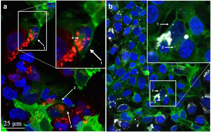
Figure 1. Intercellular Pgp-EGFP transfer.
To investigate Pgp-EGFP transfer from hCMEC/D3-MDR1-EGFP cells (Pgp-donor cells) to hCMEC/D3 cells (Pgp-recipient cells) with confocal fluorescence microscopy, Pgp-recipient cells were labeled with CMTPX (a; red) or eFluor670 (b; white) and co-cultured with an equal number of Pgp-donor cells (green). Representative images are shown. Consistent with the seeding of cells, about half of the co-culture are either red (a) or white (b) labeled Pgp-recipient cells. (a) The arrows (1–4) show the fusion protein Pgp-EGFP (in green), which was transferred to red labeled Pgp-recipient cells. Image a indicates that the Pgp-transfer to brain endothelial cells occurs by cell-to-cell contact, as in a arrow 1, a red-labeled Pgp-recipient cells (middle of magnification) that is in contact with a Pgp-donor cell (top right of magnification) receives the fusion protein Pgp-EGFP, whereas another hCMEC/D3 cell (bottom left of magnification), which is not in direct contact with the Pgp-donor cell (top right of magnification), does not receive the fusion protein. (b) In another co-culture experiment, the cytotracker CMTPX was replaced by eFluor670 to allow the combination of a further fluorescent dye (eFLUXX-ID Gold, which was used as a Pgp substrate in subsequent flow cytrometric experiments) and to confirm the results shown in a. Consistent with the results in image a, the arrows in b show a transfer of Pgp-EGFP on eFluor 670 (white) labeled Pgp-recipient cell. Fluorescent images of the co-culture in b shows DNA in blue, hCMEC/D3 cells in white and fusion protein Pgp-EGFP in green (see Fig. S1 for further explanation). Interestingly, it was observed in a and b that the Pgp-EGFP fusion protein is transferred to neighboring cells. Direct cell-to-cell contact seems to be necessary, since the fusion protein Pgp-EGFP is only transferred on labeled Pgp-recipient cells, which are in direct contact to Pgp-donor cells (arrows in a and b). In a (arrows 1–3) and in b (arrow 1) transferred Pgp-EGFP in Pgp-recipient cells is localized to the membrane in a punctuate pattern and in a (arrow 4) and b (arrow 2) also intracellularly.

