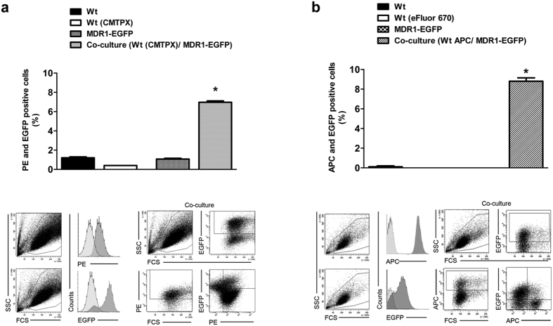
Figure 2. Flow cytometric detection of intercellular Pgp-EGFP.
Transfer of Pgp-EGFP between hCMEC/D3-MDR1-EGFP (Pgp-donor) and hCMEC/D3 (Pgp-recipient) cells was measured quantitatively by flow cytometry after 48 hours of co-cultivation. Therefore Pgp-donor cells were co-cultured with an equal amount of CMTPX (a) or eFluor670 (b) labeled Pgp-recipient cells. The extent of Pgp-EGFP transfer to the recipient cells was measured by flow cytometry of double positive PE (a, CMTPX) or APC (b, eFluor 670), and EGFP (a,b) cells. To control the double positive parameters, single cultures of unlabeled hCMEC/D3 wildtype cells, CMTPX labeled hCMEC/D3 wildtype cells (a), eFluor670 (b) and hCMEC/D3-MDR1-EGFP cells were used. Both experimental setups showed a similar extent of Pgp-EGFP transfers. The experiments were performed in triplicate with the measurement of 10,000 individual cells. Significant differences of PE (a) or APC (b) and EGFP positive cells versus control cells are indicated by asterisk (P < 0.05).

