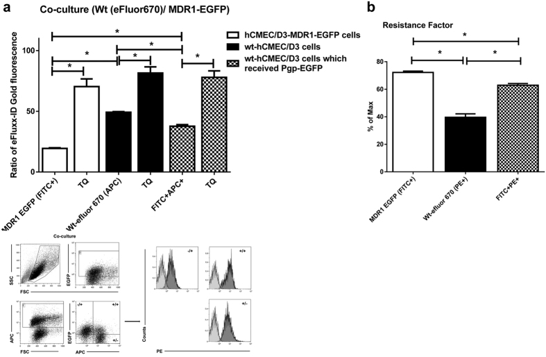
Figure 3. Intercellular Pgp transfer is functional.
(a) To characterize if transferred Pgp-EGFP in co-cultures is functional, the efflux of the Pgp substrate eFLUXX-ID Gold was measured by flow cytometry. Therefore hCMEC/D3-MDR1-EGFP cells (Pgp-donor cells) were co-cultured with an equal amount of eFluor670-labeled hCMEC/D3 cells (Pgp-recipient cells). After 48 hours of co-culturing three different cell types were identified by flow cytometry in the co-culture: 1. hCMEC/D3-MDR1-EGFP cells (open columns), 2. eFluor670-labeled wt-hCMEC/D3 cells (black columns), and 3. eFluor670-labeled wt-hCMEC/D3 cells which received the fusion protein Pgp-EGFP (hatched columns). For these three cell types present in the co-culture, alterations in Pgp efflux were indirectly measured by determining intracellular concentration of the Pgp substrate eFLUXX-ID Gold for each individual cell. The same experiments were performed in the presence of the Pgp-inhibitor tariquidar (TQ). Significant differences between the three different cell types in the co-culture in intracellular eFLUXX-ID Gold fluorescence are indicated by asterisk (P < 0.05). Tariquidar significantly increased the intracellular accumulation of eFLUXX-ID Gold and its fluorescent metabolite in all cells without inter-group differences, indicating maximal Pgp inhibition in all cell types. As expected, in the absence of tariquidar, the Pgp activity in the co-cultured hCMEC/D3-MDR1-EGFP cells (open columns) was significantly higher compared to co-cultured eFluor670- labeled wt-hCMEC/D3 cells (black columns). Transferred Pgp-EGFP was functional because eFluor670-labeled hCMEC/D3 cells, which received the fusion protein Pgp-EGFP (hatched columns), showed significantly increased Pgp activity compared to eFluor670 labeled wt-hCMEC/D3 cells (black columns) in the absence of Pgp inhibition. (b) Assessment of the multidrug resistance factor according to Huber et al.24. A 1.5-fold increase in the Pgp activity is observed in the hCMEC/D3 cells that have been co-cultured with hCMEC/D3-MDR1-EGFP cells as compared to wt-hCMEC/D3 cells. The multidrug resistance factor was calculated as described in Methods. Significant differences of intracellular eFLUXX-ID Gold fluorescence are indicated by asterisk (P < 0.05).

