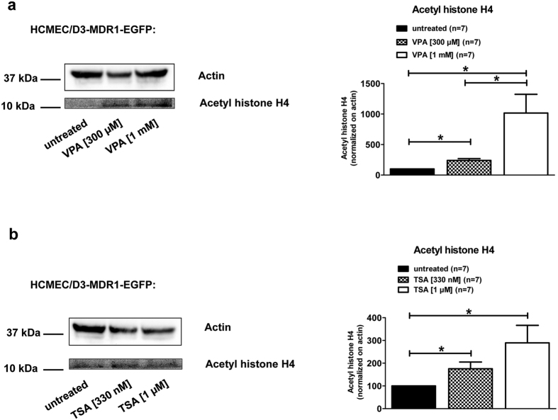
Figure 6. VPA and TSA increase acetylation of histone H4 in hCMEC/D3 cells.
Western blot studies revealed an increased acetylation of histone H4 in a concentration-dependent manner upon VPA (a; 300 μM and 1 mM) and TSA (b; 330 nM and 1 μM) treatment. In these experiments, hCMEC/D3-MDR1-EGFP cells (Pgp-donor cells) were treated for 24 hours with 300 μM or 1 mM VPA (a) or 330 nM and 1 μM of the HDAC inhibitor TSA (b). Acetyl histone H4 and actin bands of seven experiments were analyzed densitometrically and Pgp signals were normalized to actin. Significant differences of three independent experiments were analyzed densitometrically and acetylations of histone H4 were normalized versus actin.

