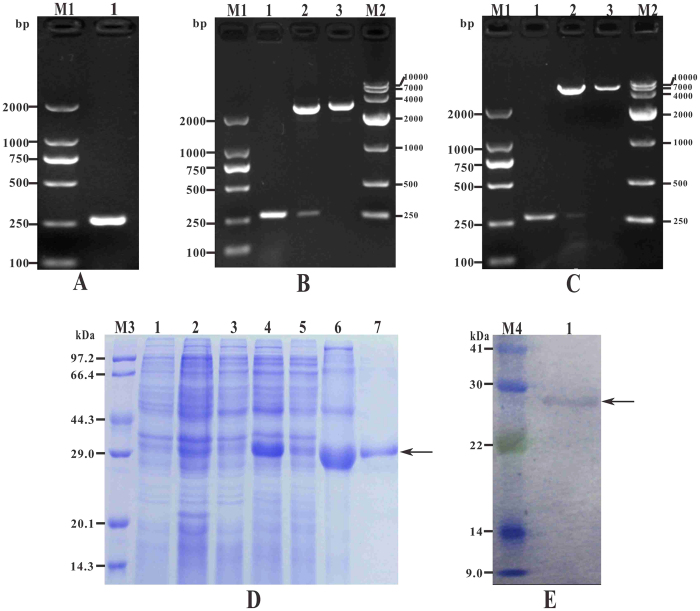
Figure 3. Molecular cloning, expression, purification and western blot analysis of rtIL-8.
(A) M1: DNA Marker (DL2000); lane 1: PCR product of tIL-8 gene with 264 bp. (B) M1: DNA Marker (DL2000); lane 1: PCR product of recombinant plasmid T-tIL-8; lane 2: digestion of T-tIL-8 with EcoR I and Xho I; lane 3: digestion of T-tIL-8 with Xho I; M2: DNA Marker (DL10000). (C) M1: DNA Marker (DL2000); lane 1: PCR product of plasmid P-tIL-8; lane 2: digestion of P-tIL-8 with EcoR I and Xho I; lane 3: digestion of P-tIL-8 with Xho I; M2: DNA Marker (DL10000). (D) M3: protein marker; lane 1 ~ 6: uninduced BL21(pET32a), induced BL21 (pET32a), uninduced BL21 (P-tIL-8), induced BL21(P-tIL-8), induced BL21(P-tIL-8) supernatant, induced BL21(P-tIL-8) sediment, lane 7: purification of recombinant rtIL-8. (E) M4: protein marker; lane 1: Specific binding between recombinant protein rtIL-8 and rabbit anti-6 × Histidine antibody.

