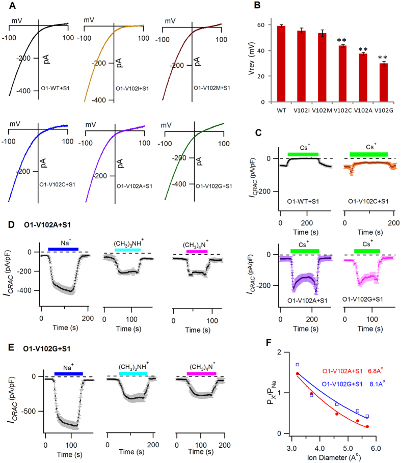
Figure 4. The Vrev, Cs+ permeability and estimated pore diameters of V102X mutants.
(A) I–V relationship (n = 6–10) of stable Ca2+ conducting ICRAC in the WT channels and V102X mutants in the presence of STIM1. (B) The bar graph summarizes the Vrev values (mean ± SEM). Compared with WT, V102C/A/G displayed significantly decreased Vrev (V102C, p = 0.00018; V102A, p = 0.0000024; V102G, p = 0.0000001). No differences were observed between V102I/M mutants and the WT channels. (C) Mean currents evaluated at −100 mV, plotted against time (n = 6 each). The extracellular solution was switched between 2 mM Ca2+ and Cs+-based DVF solution. No Cs+ conduction was observed in the WT channels. In contrast, large Cs+ currents were recorded in V102A/G channels, while a small but significant Cs+ current was detected in V102C channels. (D,E) Mean currents of DVF-Na+ or DVF-methylated ammonium cations evaluated at −100 mV, plotted against time, on V102A channels (D) and V102G channels (E) in the presence of STIM1 (n = 6–10). The extracellular solution was switched between 2 mM Ca2+ and DVF solutions containing either Na+ or the indicated organic monovalent cations. Large methylated ammonium cations mediated significant currents on both V102A and V102G mutants. (F) Px/PNa plotted against the pore diameter of each cation. The solid lines were fitted with least-squares method, following the hydrodynamic relationship. Estimated pore diameters were 6.8 Å and 8.1 Å for STIM1-activated V102A channels (red) and V102G channels (blue), respectively.

