Figure 1.

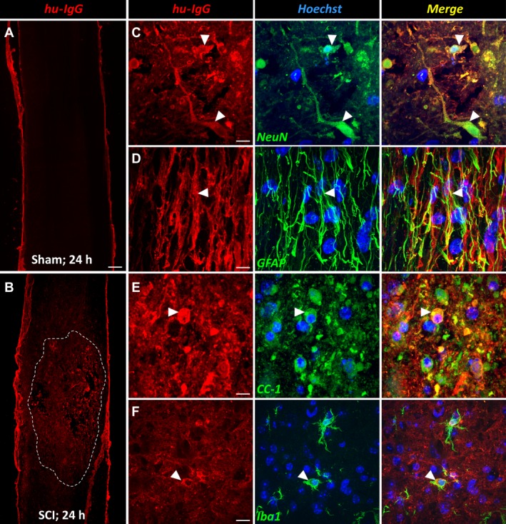
Intravenously administered immunoglobulins enter into the injured spinal cord. (A) Representative image of a sagittal spinal cord section from a sham‐operated (i.e., laminectomy only) mouse that was injected with 1 g/kg intravenous immunoglobulin (IVIg) and perfused 24 h later. IVIg (hu‐IgG staining; red) was detected near the meningeal border, but not within the spinal cord itself. (B) Mid‐sagittal section of an IVIg‐treated animal with SCI. Note that there is widespread immunoreactivity for hu‐IgG across the injured segment, which then diminishes in intensity in both the rostral and caudal directions (the damaged area is outlined by the dotted white line). (C–F) Dual‐color immunofluorescent staining showing co‐localization between hu‐IgG and NeuN+ neurons (C, arrowheads), GFAP + astrocytes (D, arrowhead), CC1+ oligodendrocytes (E, arrowhead), and Iba1+ microglia (F, arrowhead). Scale bar: A, B (in A): 200 μm; C: 15 μm; D: 11 μm; E: 13 μm; F: 22 μm. GFAP, glial fibrillary acidic protein; SCI, spinal cord injury.
