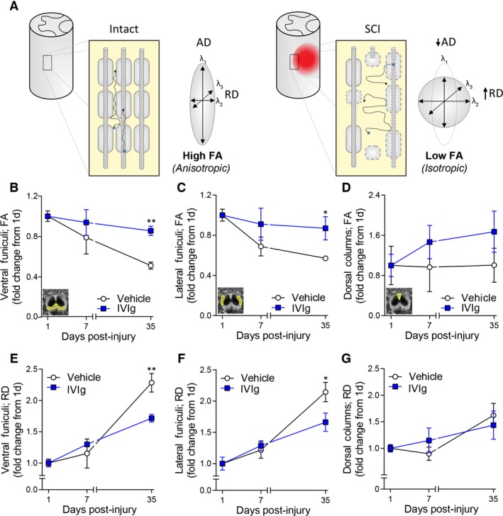
Figure 3.

IVIg treatment improves diffusion tensor imaging (DTI) measures. (A) Schematic diagram explaining anisotropic and isotropic diffusion in spinal white matter based on DTI principles. Blue dots indicate water molecules and their net trajectory of diffusion (arrows) as influenced by the cytoarchitecture of the spinal cord white matter under homeostatic and injured conditions. Diffusion of molecules in the spinal white matter is normally highly restricted (anisotropic) by anatomical barriers such as axonal membranes and myelin sheaths (left). Axonal damage and loss of myelin following spinal cord injury (SCI) (right) disrupts these barriers, leading to less directional (isotropic) diffusion, lower fractional anisotropy (FA), and higher radial diffusivity (RD). (B–D) Temporal change in FA at the lesion epicenter for the ventral funiculi (VF; B), lateral funiculi (LF; C), and the dorsal columns (DCs; D). Note the deterioration of FA values in the VF and LF of vehicle‐treated mice between 1 and 35 days post‐SCI, and that IVIg treatment attenuated this pathological change in FA values for these ROIs. No significant temporal change in FA values was observed for the DCs in either experimental group. (E–G): Note the increase in RD between 1 and 35 days post‐SCI at the lesion epicenter for both the VF (E) and LF (F). IVIg treatment significantly counteracted these SCI‐induced increases in RD. As with FA, no significant time or treatment effect was observed for RD values in the DC area (G). All data points represent mean ± SEM; n = 5–7 per experimental group; *P < 0.05; **P < 0.01; two‐way repeated measures ANOVA with Bonferroni post hoc test. ANOVA, analysis of variance.
