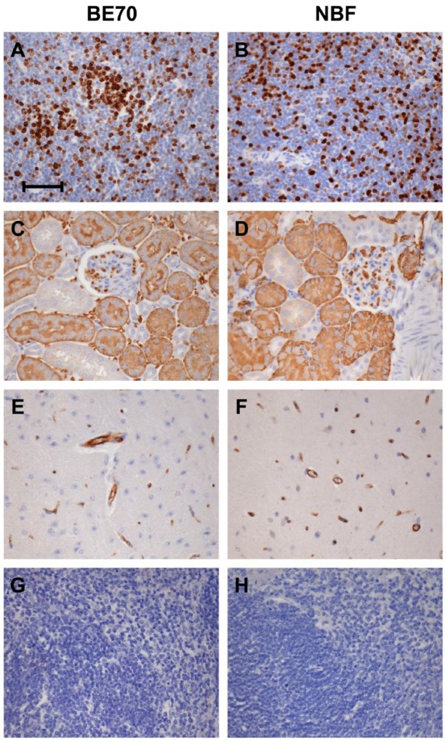
Figure 3.
Immunohistochemical staining between buffered ethanol 70% (BE70) and neutral-buffered formalin (NBF) fixation. Immunohistochemistry staining with anti-Ki-67 in the mouse spleen (A and B), anti-aquaporin 1 (AQP1) in the mouse kidney (C and D), anti-CD31 in the mouse brain (E and F), rabbit immunoglobulin G (IgG) isotype control (G), and no primary antibody control (H). Scale bar, 50 µm.

