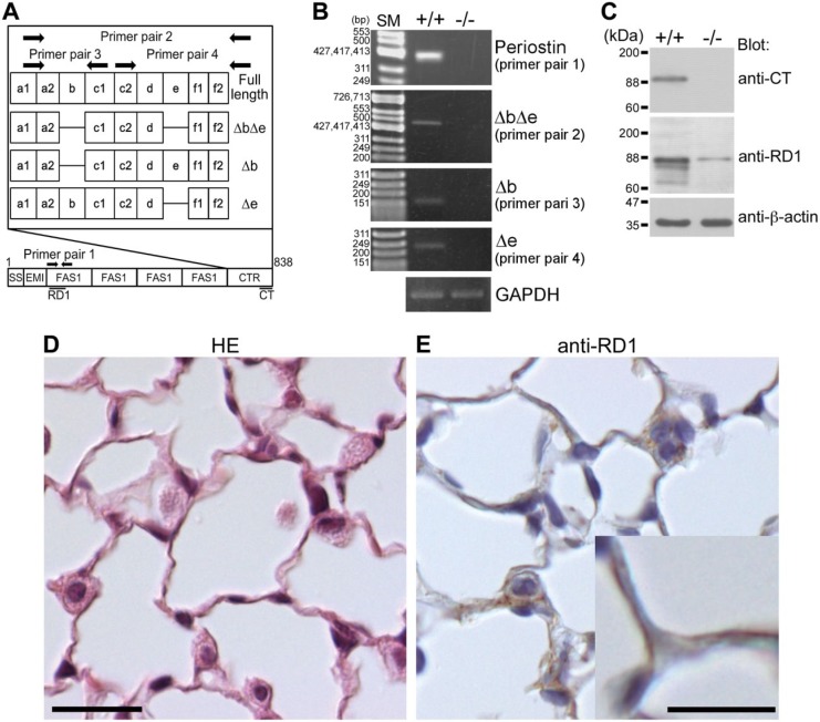
Figure 1.
Expression of periostin in the lungs of wild-type mice. (A) Pattern diagrams of the periostin splice variant forms, primer pairs, and antibody recognition sites. SS, EMI, FAS1, and CTR mean signal sequence, EMI (EMILIN) domain, Fasciclin 1 domain, and C-terminal region, respectively. Periostin antibody recognition sites are indicated as black lines. (B) Expression of the periostin alternatively spliced variant form ΔbΔe in lung tissue from 8-week-old wild-type mice. Details on the primer pairs 1, 2, and 3 are described in the “Materials and Methods” section. GAPDH is shown as an internal control. +/+ indicates wild type and −/− indicates negative control (periostin-null mouse). SM means size marker. (C) Expression of periostin protein in lung tissue. Tissue lysates from the lungs of 8-week-old wild-type and periostin-null mice were subjected to SDS-PAGE. Proteins were visualized by blotting with anti-CT, anti-RD1, and anti-β-actin antibodies. A band corresponding to intact periostin is located at the 90-kDa position and those of cleaved periostin at approximately 84 and 74 kDa. +/+ indicates wild type and −/− indicates negative control from periostin-null mouse. The band located at the 90-kDa position in the −/− lane indicates a nonspecific signal. (D) Histological section of a lung alveolus of an 8-week-old wild-type mouse. The paraffin section was stained with hematoxylin–eosin (HE). (E) Localization of periostin protein in a lung alveolus from an 8-week-old wild-type mouse. The paraffin section was stained with anti-RD1 antibody, which was then counterstained with hematoxylin. Periostin is localized in the alveolar walls and in type II pneumocytes. Scale D = 20 µm; Scale E = 20 µm (high magnification = 10 µm).

