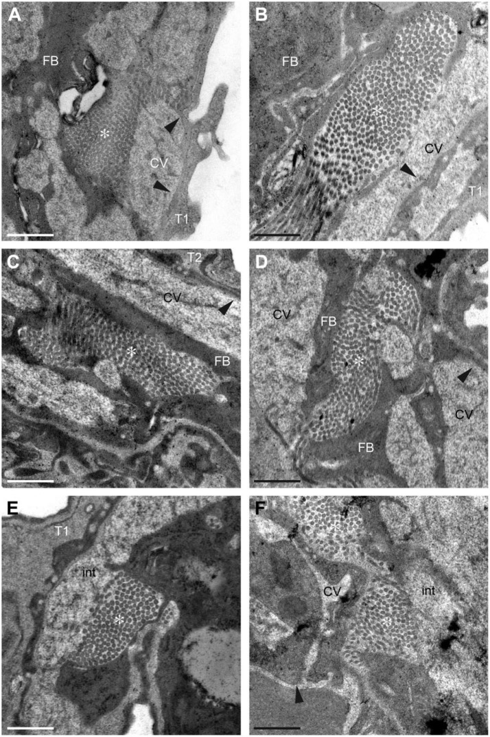
Figure 4.
Transmission electron microscopy (TEM) analysis of wild-type and periostin-null lung tissue. (A–E) Electron micrographs of lungs from an 11-week-old wild-type (A, C, E) and from a periostin-null (B, D, F) mice. Arrowheads indicate vessel walls, and * indicates collagen fibrils. Scale = 500 nm. Abbreviations: T1, type I pneumocyte; T2, type II pneumocyte; FB, fibroblast; CVs, capillary vessels; int, interstitium.

