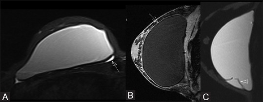
Figure 10 (A-C).

MRI of intact implants. The axial T2-weighted TIRM image (A) of the breast shows minimal normal periprosthetic fluid collection (closed arrow). The low signal intensity capsule (open arrow) can be very well appreciated in the sagittal T2-weighted image (B). Sagittal T2-weighted TIRM image of breast demonstrating a silicone gel implant with normal radial folds (arrowhead) (C). Periprosthetic fluid and radial folds are not in themselves indicative of rupture
