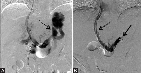
Figure 5 (A and B).

12 mm × 9 mm AVP used to embolize the coronary vein concomitantly with TIPS revision due to gastric fundal varices, with resultant improved portal flows post-embolization. (A) Pre-deployment shows prominent porto-systemic shunting through the coronary vein (dotted arrow) and (B) post-deployment of the AVP (arrow) occluding the coronary vein with improved flow through the TIPS shunt (double arrow)
