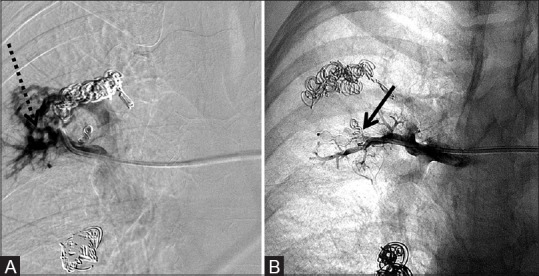
Figure 9 (A and B).

6 mm × 6 mm AVP deployed into the right upper lobe pulmonary artery main feeding trunk to occlude a pulmonary arteriovenous malformation after failure of attempted coil embolization twice with 3 mm × 3 cm microcoils. (A) Pre-deployment shows persistent vascular malformation (dotted arrow) and (B) post-deployment of the AVP (arrow) with lack of filling of the malformation
